The Journal of Targeted Therapies in Cancer
- 2016 August
- Volume 5
- Issue 4
Advances in Image-Guided Oncologic Treatment
Advances in the computerization of the imaging, blood flow, and tumor measures of exact volume and vessel density are now less operator dependent. That provides for an accurate and repeatable diagnosis, and a means to follow the individual patient’s unique pattern of cancer development, progress, and response to treatment.
Abstract
Three-dimensional sonography and power Doppler angiography are techniques that contribute new morphologic parameters and noninvasive functional tumoral angiogenic markers for evaluation and treatment follow up of hyperplastic diseases and prostate cancers. Accuracy in assessing bladder breast, skin/ melanoma, liver, endometrial, and thyroid malignancies have been documented. Diagnostic ultrasound is a viable means to assess these lesions and can be performed in the office setting accurately and rapidly due to the high resolution and low cost of today’s sonographic equipment. This diagnostic technology requires extensive experience and training in interpreting the images. However, advances in the computerization of the imaging, blood flow, and tumor measures of exact volume and vessel density are now less operator dependent. That provides for an accurate and repeatable diagnosis, and a means to follow the individual patient’s unique pattern of cancer development, progress, and response to treatment.
Introduction
Summer means more adults will seek reassurance about pigmented lesions and patients with a previous dermal or other malignancy will need a diagnosis on any palpable lesions that may be subdermal in location and, thus, invisible to the spatially restricted human eye. To assess the condition of these lesions, clinicians can look to biopsy or the use of gadolinium- based contrast agentsintravenous drugs to enhance the quality of magnetic resonance imaging (MRI) or magnetic resonance angiography. However, patients are concerned about possible side effects associated with these techniques.
Diagnostic ultrasound is a viable means to assess bladder1breast2, skin/melanoma3, liver, endometrial4, prostate, and thyroid malignancies and can be performed in the office setting accurately and rapidly due to the high resolution and low cost of today’s sonographic equipment. This diagnostic technology requires extensive experience and training in interpreting the images. However, advances in the computerization of the imaging, blood ow, and tumor measures of exact volume and vessel density are now less operator dependent. That provides for an accurate and repeatable diagnosis, and a means to follow the individual patient’s unique pattern of cancer development, progress, and response to treatment. Recent technological advances also make these procedures available to much broader clinical application, without requiring years of unique training and experience, for example, with diagnoses of cystic versus solid lesions. These advances are applicable beyond prostate cancer, the example used here, because it has been shown to be reproducible over the last 20 years with pathologic confirmation of the findings of the various imaging modalities. For the clinician new to the use of these technologies, it must be emphasized that initial readings will be difficult to interpret and may contain many confusing artifacts. It is recommended that findings should be confirmed with all pertinent imaging modalities.
Prostate Cancer
One man in 6 will be diagnosed with this disease in his lifetime. It is, at the same time, the second biggest cancer killer in men, with an esti- mated 29,720 deaths in 2015 in the United States. Like other cancers in the past, our understanding of the science of prostate cancer has changed tremendously during the last 10 years. Pre-malignant conditions have been described leading to an extremely active search for genomic signatures of prostate cell transformation. Cohort studies are ongoing. The diagnosis of prostate cancer has become more sophisticated with the introduction of newer criteria, outside of the classi- cal Gleason classi cation, that could predict an individual’s tumor aggressiveness, with the hope of better and more personalized tailored therapeutic strategies.53D imaging may not detect regional and distant lymph nodes, so MRI remains the gold standard for nodal staging.
Among potential tailored treatments, active surveillance is more widely accepted and fewer patients are choosing to undergo an unnecessary prostatectomy, which has been on the rise in the last decade because of widely increased use of prostate-specific antigen (PSA) screening. This is a reason for some real concern, both in term of individual risk as well as for the economy of cancer. Because of the potential for overuse and overtreatment, the United States Preventive Services Task Force (USPSTF) recommended against routine PSA screening. Given the resulting shift towards fewer PSA tests and its low accuracy, we are seeing a rise in non-invasive screening with Doppler ultrasound and MRI.
Three-dimensional Doppler ultrasound with Dynamic Contrast Enhanced MRI are the gold standards by which cancers are initially diag- nosed and serially followed after treatment. The percentage of malignant vessels can be quantified and re-evaluated in the identical tumor volume as serial follow ups. Since vessel mapping is possible, embolic treatments may be used.
Three-dimensional sonography can demonstrate the prostate capsule more accurately than MRI because the resolution is 100 microns at 18MHz. The examination takes about 10 minutes and the probes are automated meaning that this is less operator dependent than other sonographic procedures. Vessel density index (VI) imaging is performed on the data set at an inde- pendent workstation and comparison made with prior examinations if available.
Three-dimensional power Doppler indices vary according to the tumor stage, the histologic grade, capsular disruption, and lymph node metastases. Histologic grade has been studied with this technology and the following approximation has proven useful: low- grade tumor (Gleason 3+2, 3+3) has VI <5%, medium-grade cancer (Gleason 3+4) has VI 5-9% and high-grade malignancies (Gleason 4+4, 4+5, 5+5) have VI >9%. This does not exactly correlate with histologic Gleason grading since this is a current functional measure while the microscopy is purely anatomical and may not represent current aggressive potential.
Skin Cancer
Major changes in the incidence, diagnosis, and treatment of skin cancer highlight a singular need for an up-to-date source of diagnosis and minimally invasive therapy of dermal tumors. The occurrence of melanoma is now at a median age of 28 years and the incidence is increasing rapidly. This means patients in their 30s are developing cancer that may be missed by current screening technologies of dermoscopy, confocal microscopy, and near infrared optical devices. Delays in treatment due to misdiagnosis have led to lawsuits. Earlier detection of tumors means smaller lesions are being discovered and focal nonsurgical treatment may be preferred to standard operative modalities with potential long-term postoperative side effects.
Medical imaging can map the arteries, veins, and nerves providing preoperative landmarks reducing postoperative bleeding and avoiding nerve damage. Tumors of low aggressive potential may be treated medically and followed by interval scans or locally reduced by radiation or laser ablation. Biopsies of certain abnormalities may be averted or postponed.
Dermal sonography has been used since 1980 and complements optical media such as dermoscopy, con-focal microscopy, optical coherence tomography (OCT), dermal CT, and MRI scanning. Collectively, these media help the clinician diagnose, stage, and grade cancer. Dermal sonography evaluates the extent of certain benign disorders such that biopsy may be limited or avoided. This is the case when part of a lesion is cancer and the adjacent areas of the clinical lesion are benign due to brosis, in ammation, hyperplasia, and immune-cell activity.
Clinical diagnosis of nonmelanoma skin cancer (NMSC) is accurate, however, the depth of a tumor is unknown. Imaging informs the surgeon whether the surgery will be limited or extensive and may require skin graft or cartilage replacement. Clinical diagnosis of malignant melanoma is 54% accurate by histology and 20% accurate by nonmicroscopic clinical modali- ties. Many benign pigmented lesions are removed pre- ventively. There is 1 melanoma found per 33,000 nevi. Ultrasound screening of pigmented lesions is highly accurate and well tolerated. MRI studies are currently sensitive but not speci c for screening. Dermoscopy and other optical technologies are complementary. Pa- thologists now preview clinical pictures of a suspect lesion before they nalize readings due to the inherent variability of interpretation. The finding of a subclinical metastatic focus near the lesion provided by the newer ultrasound and spectral technologies facilitates histologic interpretation.
Lymph Node Disease
Lymph node assessment is possible at the same time. Under sonographic guidance, biopsies may be obtained. Sonographic criteria for malignancy are published elsewhere. Image guidance of enlarged nodes can distinguish between active tumor and necrotic areas, and thus diminishing the necessity for repeated aspirations for indeterminate findings.
Endometrial/Cervical Cancer
While surgery is the current standard, there have been documented cases of antioxidant treatment successfully regressing these over periods up to ten years. As in other cases, neovascular response correlates with treatment effect.
Thyroid Cancer
Vascular lesions may be treated with ablative technologies with significant reduction in vessel density correlating with improvement.
Hepatic Cancer/Metastatic Diseases
Reduction in tumor neovascularity correlates with improvement. Indeterminate areas may be imaged with contrast-enhanced ultrasound as an off-label procedure that is proving highly accurate in European medical centers.
Breast Cancer
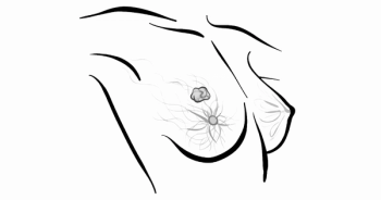
High tumor vessel density correlates with greater aggression. Axillary and mediastinal imaging can document lymphadenopathy. Abdominal scans simultaneously performed may detect ascites and metastases to the liver, periaortic nodes, and pelvic organs. Response to neoadjuvant chemotherapy may be assessed using MRI, computed tomography (CT), mammography, PET/CT, and ultrasound. The new technology of ultrasound elastography, which can assess tumor stiffness, predicts response to treatment accurately and may indicate better therapeutic strate- gies on a timelier basis.6
Image-Guided Biopsy and Treatment
New computer programs use nanotechnology and cybernetic modalities for accurate image-guided biopsy and treatment options. The physician can employ 3D sonography with Doppler, manually targeting the area of highest tumor neovascularity. This is critical because only part of a mass may be cancerous and can be missed on nontargeted punch biopsies. The marriage of fusion of MRI with ultrasound permits image-guided biopsies that spare the adjacent neurovascular bundles. The same technology allows customized ultrasound or MRI guided biopsies to be performed under local anesthesia. Immediate cytologic confirmation of tumor cells permit the withdrawal of the biopsy needle and insertion of a laser ber or cryogenic probe immediately treating the proven tumor.7MRI thermocoupled sensors prevent overheating of the adjacent nerves and sensitive tissues. Following ablation, the zone of destruction is con rmed with Doppler, contrast ultrasound, or dynamic contrast-enhanced MRI. In amatory lesions that are deeply seated may be approached by robotic image-guided subdermal injections or targeted biop- sies if necessary. This outpatient procedure allows the patient to return to work immediately. Radiofrequency thermoprobes with automatic temperature cutoffs prevent thermal skin damage. Similar user-friendly and cost-effective modalities may replace other thera- pies in the near future. At the 2016 ASLMS meeting in Boston, cutaneous melanoma with intransit metastases were successively treated by laser technologies.
Summary
3D sonography and power Doppler angiography are techniques that contribute new morphologic param- eters and noninvasive functional tumoral angiogenic markers for evaluation and treatment follow up of hyperplastic diseases and prostate cancers. 3D PDS has been proven to be of value in identifying extraprostatic tumor extension. It is clinically useful in the detection and grading of breast cancer and endometrial carcinoma. Density analysis of malignant melanoma serves as a guide to biopsy of a skin lesion, treatment in transit lesions by image-guided technologies, and follow up of metastatic foci at later intervals. It has been applied in the characterization of bladder tumors which are occasionally imaged during the course of a TRUS. Abnormal disoriented irregular vessels in prostate cancer are present in 69% of cases with Gleason 7 or greater tumors. External beam radical radiotherapy remains the primary treatment for patients with locally advanced or high grade tumors in the United States as is associated with a 5-year failure rate of 35% with Gleason scores of 6 or higher. The biochemical failure rate predicting tumor recurrence is less reliable than thought and may be replaced by other functional parameters such as DCE MRI and tumor Vascular Endothelial Growth Factor (VEGF) expression. The addition of in vivo real time volume density measurements of tumor neovascularity may prove to be of value in determining the risk of tumor recurrence noninvasively.8
Residual cancer burden scoring could provide better treatment options since the treatment response for evaluation of neoadjuvant chemotherapy needs a more comprehensive and authoritative standard than is currently available.9,10
Dermal tumor depth can be accurately assessed, thus shortening MOHS treatments. Cartilage invasion and aberrant nerves or arteries may be preoperatively as- sessed. Subcutaneous masses such as cysts or foreign bodies may be diagnosed. Given the increasing use of biologic agents, with their accompanying adverse effect of lymphoma, advanced sonography techniques can assure patients that a new palpable lesion is indeed a benign lipoma or cyst, which can be readily differentiated from a metastatic node or lymphocytic cancer. The more oncologists, dermatologists, and plastic surgeons who use the imaging capacities of sonography, the more they will find its attributes essential to the modern practice of skin disorder therapies.
References:
- Bard R, Cantisani V, Barentsz J. Vascular imaging of bladder cancer. J Radiol 2010;91(10):1479.
- Bard R. Vascular imaging of cancer in the dense breast. 60th Journees Francaises de Radiologie, Paris. 2012, p 416.
- Bard R. Image guided cancer treatment. Advances in Medical and Surgical Dermatology, 15th Annual Mt Sinai Winter Symposium 2012 New York.
- Merce L, Alcazar J, Lopez C et al. Clinical usefulness of 3 dimensional sonography and power Doppler angiography for the diagnosis of endometrial carcinoma. J Ultrasound Med. 2007;26(10):1279-1287.
- Jing M, Wen C, Zi L, et al. Early evaluation of relative changes in tumor sti ness by elastography predicts response. J Ultrasound Med. 2016; 35(8):1619-1627. doi: 10.7863/ultra.15.08052..
- Öbek C, Doğanca T, Erdal S, Erdoğan S, Durak H. Core needle biopsy length a ects grading and staging. J Urol. 2012;187(6):2051-2055. doi: 10.1016/j. juro.2012.01.075.
- Zalesky M, Urban M, Smerhovský Z, Zachoval R, Lukes M, Heracek J. Value of power Doppler sonography with 3D reconstruction in preoperative diagnostics of extraprostatic tumor extension in clinically localized prostate cancer. Int J Urol. 2008;15(1):68-75. doi: 10.1111/j.1442-2042.2007.01926.x.
- Symmans WF, Peintinger F, Hatzis C, et al. Measurement of residual breast cancer burden to predict survival after neoadjuvant chemotherapy. J Clin Oncol. 2007;25(28):4414-4422.
- Bard RL, Fütterer JJ, Sperling D (eds). Image Guided Prostate Cancer Treatments. [1st edition.] Berlin, Heidelberg, Germany: Spring-Verlag; 2014.
Articles in this issue
over 9 years ago
Pembrolizumab Shows Potential in Advanced Thyroid Cancerover 9 years ago
MABp1 Improves Symptoms in Phase III CRC Studyover 9 years ago
Redefining Multiple Myeloma Diagnosis and Management


