Overview and Diagnosis of Differentiated Thyroid Cancer
Lori Wirth, MD, provides an overview and diagnosis consideration of differentiated thyroid cancer and its related subtypes.
Episodes in this series

Lori Wirth, MD: I’m Lori Wirth, and I’m a medical oncologist at Massachusetts General Hospital in Boston, Massachusetts.
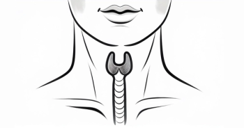
For a brief overview of differentiated thyroid cancer: It is a spectrum of various thyroid cancers that derive from the follicular cells in the thyroid gland. There are several subtypes; the most common subtype, a papillary thyroid cancer, follicular thyroid cancer, Hürthle cell carcinoma, and poorly differentiated thyroid cancers. All these subtypes are referred to as differentiated thyroid cancer to distinguish them from anaplastic thyroid cancer or medullary thyroid cancer.
Overall, in the United States, we see approximately 45,000 patients every year who are newly diagnosed with differentiated thyroid cancer. In terms of the prognosis, most patients with differentiated thyroid cancer will do quite well. Most patients will be cured with treatment involving surgery plus or minus radioactive iodine. There is, however, a subset of patients who do not do well. In the United States we see approximately 2200 deaths per year from thyroid cancer.
What are the risk factors for thyroid cancer? We think most thyroid cancers don’t have identified risk factors thus far that we’ve been able to figure out. Thyroid cancer is more common in women; we see 3 times the diagnosis of thyroid cancer in woman that we do in men. We do know that radiation exposure, particularly in childhood, can cause thyroid cancer. For example, that’s been well described by the Chernobyl incident. Then there is a small subset of patients with differentiated thyroid cancer that have the cancer in relation to an inherited familial syndrome, such as Cowden [syndrome] or the DICER1 germline mutation.
Differentiated thyroid cancer is often diagnosed when it is found incidentally when a patient has imaging for another problem; for example a chest CT scan may show in the upper cuts a mass arising in the thyroid gland, or a PET [positron emission tomography] or CT scan can identify an FDG [fluorodeoxyglucose]–avid nodule in the thyroid gland. However, patients can also present to their clinicians complaining of a neck mass that they’ve noticed, or it can also be found on a good physical examination by a primary care physician or another treating clinician.
Once that neck mass is identified, the standard process is for patients to have an ultrasound to characterize the appearance and size of the nodule. If there are risk factors for malignancy, typically the next diagnostic intervention is a fine-needle aspiration and cytology. Molecular diagnostic can be quite helpful in the setting of an indeterminate fine-needle aspiration. If there are cells that appear atypical or suspicious but not entirely diagnostic for thyroid cancer, there are a couple of molecular diagnostic tests that are available on the market that can give a good indication for whether that indeterminate nodule is malignant.
Transcript edited for clarity.


