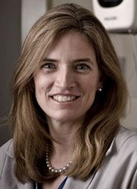
Friedewald Highlights Impact of Tomosynthesis on Breast Cancer Screening
Sarah M. Friedewald, MD

Sarah M. Friedewald, MD
While tomosynthesis has been described by the oncology community as one of the biggest improvements in breast cancer screening, the technology that renders 3D-like images of the breast does carry limitations. The implementation of tomosynthesis, however, has allowed for more accurate detection of breast cancersespecially in patients with dense breasts, explains Sarah M. Friedewald, MD, division chief, Breast and Women’s Imaging, and medical director, Lynn Sage Comprehensive Breast Cancer Center, Northwestern Memorial Hospital.
Additionally, tomosynthesis has proven to be useful in an era where radiologists are challenged by the ever-changing recommendations of when breast cancer screening should begin and the frequency of screening, she noted during a presentation at the 2016 Lynn Sage Breast Cancer Symposium.
Tomosynthesis makes the malignancy more apparent. Friedewald described tomosynthesis screening as visually “peeling back” the breast tissue and being able to dissect the image slice by slice. The number of images a tomosynthesis machine takes is based on the individual manufacturer recommendations, explained Friedewald, and not a radiologist’s decision. “For example, if a breast has a 5% compression, the tomosynthesis scan might result in 54 images; but not to be confused, there’s not 54 images of the breast taken for us to look at.”
Tomosynthesis received FDA approval in 2011. At the end of 2015, there was a 20% adoption rate in the United States, said Friedewald. In January 2015, Medicare approved it for use. As of June 2016, Cigna, one of the country’s largest insurers, approved it as a mammogram option because of the NCCN guidelines, said Friedewald. All of these factors led tomosynthesis to be adopted much more quickly than the adoption rate of digital over film.
What is commonly referred to as tomosynthesis screening is not actually just a 3D image alone, explained Friedewald, but actually combined data using both 2D and 3D images. The radiologist receives 3 sets of data from a tomosynthesis scanthe raw data, which are not used by most radiologists, a 2D scan, and the reconstituted 3D images, said Friedewald. The 3D images can be further dissected into 1 mm slices to distinguish abnormalities, she said.
There are numerous clinical data supporting the use of tomosynthesis, said Friedewald. It has been evaluated in 2 radiologist reader studies. In one study, every radiologist reading improved when tomosynthesis was added. These were the studies presented to the FDA that led to the approval of tomosynthesis.
In a retrospective analysis of 454,850 examinations at 13 different institutional sites that were early adopters of tomosynthesis, conducted by Friedewald and her team, there was a decrease in the overall rate of recall by a relative difference of 15%.1There was also an increase of 41% in invasive cancer detection.
In the 13 sites, most moved from a decrease in recall rate to an increase in cancer detection with tomosythesis use, noted Friedewald. Only 2 sites went in the opposite direction. In one, the sample size was too small to be statistically significant, she concluded. The other site only had 6 months of use with tomosythesis, and Friedewald explained that there’s a learning curve with using tomosynthesis that may have contributed to that outcome.
Friedewald provided real life imaging examples of the benefits of tomosynthesis. In one example, tomosynthesis detected a bilateral invasive carcinoma that 2D imaging had missed. “If she had only had the 2D image, she would’ve been dismissed home and would come back in a year.” In another case, tomosynthesis differentiated that what appeared to be a speculated tissue was actually a blood vessel turn. With a 2D image, that may have led to further biopsy, noted Friedewald.
“Overall, with tomosynthesis, there’s an increase in cancer detection and a simultaneous decrease in recall,” said Friedewald of the technology’s major benefits.
Friedewald admitted that there are limitations to tomosynthesis. Of particular note, there are significant variabilities based on breast densities, said Friedewald. This is important, explained Friedewald, because women defined as having dense breasts can have either heterogeneously dense breast tissue or extreme dense breast tissue.
“Still more studies need to be done to recognize the benefits in each of those categories,” she argued.
“It isn’t perfect. A lot of people think tomosynthesis is the answer and that they don’t need to do anything else. That’s simply not true,” cautioned Friedewald. “There are some things
it misses but it’s still a big improvement over 2D alone.”
Friedewald provided examples of where an ultrasound after tomosynthesis imaging provided additional information the new technology had missed. In one case a large mass detected a year after the initial tomosynthesis screening showed no cause for concern.
Friedewald also cautioned against a limited use of tomosynthesis, for example 3D imaging with MLO view only, as more views may be needed to detect the widest range of abnormalities. “Some cancers are still only seen in one projection,” explained Friedewald.
Also, for all sites reporting on tomosynthesis use, there was no change in ductal carcinoma in situ detection with tomosynthesis, said Friedewald.
Perhaps the biggest plus for tomosynthesis is the role it plays in boosting the recommendation for annual mammograms, an area of continued controversy in the breast cancer care community. “With tomosynthesis, we are really able to drop that false-positive rate, which is part of the major concern with annual cancer screenings,” concluded Friedewald.
Reference:
Friedewald SM, Rafferty EA, Rose SL, et al. Breast cancer screening using tomosynthesis in combination with digital mammography.JAMA.2014;311(24):2499-2507.
In traditional, 2D mammography, Friedewald explained, there’s a high sensitivity on identifying invasive abnormalities in fatty breasts, but that sensitivity drops significantly in women with dense breast tissue, who account for half of the screening population. “2D mammography can hide an abnormality and it can also make it appear there’s a lesion there when it doesn’t really exist, resulting in false-negatives and recall,” said Friedewald.