First Patient Enrolled in ON-SITE Study of AI-Based Image Analysis for Lung Cancer Detection
The ON-SITE study has enrolled its first patient to evaluate an AI-based image analysis module for lung cancer.


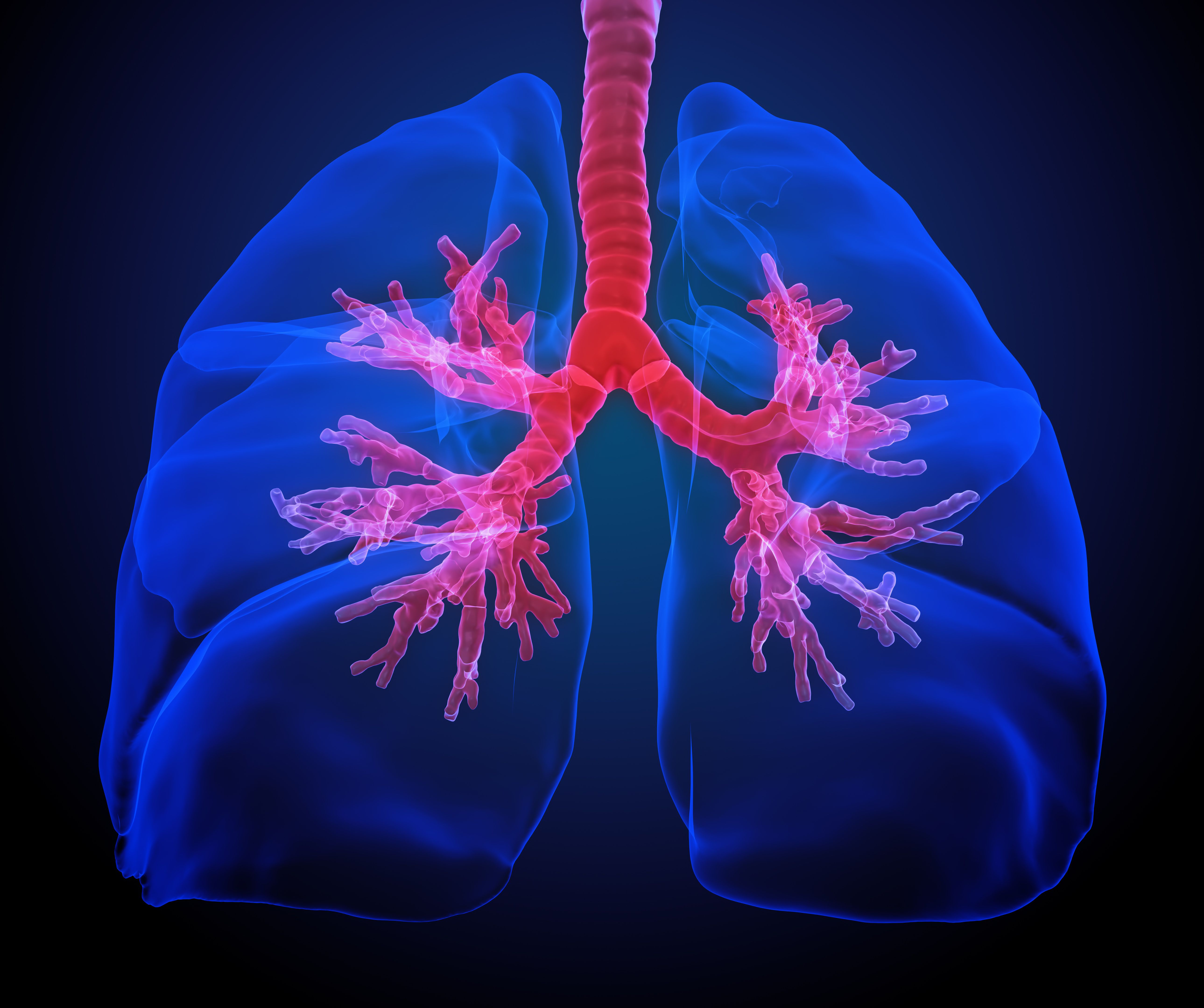
3D artistic rendering of human lungs: © Mopic - stock.adobe.com
The ON-SITE study has enrolled its first patient.1 This multicenter investigation will explore the effectiveness of an artificial intelligence (AI)-based image analysis module for lung cancer detection.1,2
The AI-based image analysis module for lung cancer is being evaluated by researchers at the University of North Carolina.
By combining stimulated Raman histology with AI, the study aims to enhance the speed and accuracy of lung cancer diagnosis during bronchoscopy. The NIO® Laser Imaging System offers rapid imaging of fresh tissue biopsies within the treatment room. The design of the system eliminates the need for staining or sectioning processes. This allows the existing operating room staff to handle sample preparation efficiently.2
"[Rapid-on-site tissue evaluation (ROSE)] requires that a cytologist or highly trained cytotechnician be physically present in the procedure room, and thus it is not available at many centers performing lung biopsy due to resource limitations," said Jason Akulian, MD, director of interventional pulmonology at the University of North Carolina at Chapel Hill, in a press release. "We are excited by the NIO's potential to extend the benefits of ROSE to the proceduralist when the service is not available."
Lung cancer remains the leading cause of cancer-related mortality in the US, prompting the implementation of extensive screening programs for high-risk populations. These efforts have led to the identification of approximately 3.1 million new primary lung nodules annually.
Though there have been significant advancements in minimally invasive biopsy technologies, obtaining sufficient tissue for accurate biomarker analysis and treatment decisions continues to be a challenge. Consequently, current bronchoscopy guidelines advocate for ROSE to enhance the adequacy and diagnostic yield of lung biopsies.
The system's NIO® Slides also allow for easy retrieval of samples for further analysis. With inherently digital imaging capabilities, NIO® images can be shared almost in real time. The ON-SITE study is dedicated to creating and validating an AI-driven image analysis module for the NIO® Laser Imaging System, aiming to aid physicians in detecting cancer in bronchoscopic lung biopsies, especially in cases where ROSE is not available.
"Artificial intelligence aiding healthcare may seem utopic, but the future is coming. While still investigational, the promise of fast, in-room, accurate identification of tissue that is suspicious for cancer has the potential to ultimately lead to improved outcomes, a beneficial cost/benefit profile, and personalized treatments," said Gustavo Cumbo-Nacheli, MD, pulmonologist at Corewell Health, in a press release.
"Enrolling the first patient in the ON-SITE study is an important milestone for Invenio, as we aim to develop the first FDA-cleared AI to identify cell/tissue morphology suspicious for cancer in lung biopsies," said Jay Trautman, PhD, co-founder and chief executive officer of Invenio Imaging, in a press release. "Near real-time image analysis on the NIO® Laser Imaging System completes the end-to-end solution for streamlined intraoperative histology."