Singal Discusses the Importance of Biopsy Testing When Considering HCC Treatment
During a Targeted Oncology™ Case-Based Roundtable™ event, Amit Singal, MD, discussed next steps after diagnosing a patient with metastatic hepatocellular carcinoma.


Amit Singal, MD
Medical Director of the Liver Tumor Program
Chief of Hepatology
Harold C. Simmons Comprehensive Cancer Center
UT Southwestern Medical Center
Dallas, TX

CASE SUMMARY
- A 77-year-old White woman presented to her primary care physician complaining of abdominal pain and fatigue.
- She had a history of cirrhosis due to heavy alcohol use; Crohn disease, controlled with infliximab (Avsola); and a history of variceal bleeding, with banding 2 months ago.
- Her ECOG performance status scale score was 1.
- A CT scan of her chest, abdomen, and pelvis with triphasic liver evaluation showed a 4.5-cm LI-RADS (LR) 5 hepatic mass in the right lobe plus metastatic disease in the lung.
- She had a Child-Pugh score of A and α-fetoprotein (AFP) level of 380 ng/mL.
Targeted Oncology: Is an MRI needed for a diagnosis of hepatocellular carcinoma (HCC), or is a CT scan sufficient?
SINGAL: This question of CT scan vs MRI is a valid one, as both a CT scan and MRI are acceptable. The MRI does have a higher sensitivity than the CT scan, so if you can get either one, I personally think a good-quality MRI is better than a good-quality CT scan. That being said, if you have a good-quality CT scan and you have characteristic appearance, this does not mean you have to get an MRI. This does not mean you have to get a biopsy. The positive predictive value for that imaging appearance of LR 5 is about 95% to 97%.
How important is AFP in diagnosing HCC?
The AFP levels used to be part of the diagnostic criteria. The cutoff used to be 200 vs 400 ng/mL, but these were the criteria that would be considered for diagnosis of HCC. More recently the guidelines have removed that from the diagnostic criteria, given both poor sensitivity and specificity.1 The AFP sensitivity, in terms of diagnostic criteria, is only about 50%. We see many [cases of patients with] HCC where you can even have normal AFP levels, despite having metastatic disease or vascular invasion. We also know that AFP can be elevated in the setting of mixed tumors or even some cholangiocarcinoma itself, so AFP is not part of the diagnostic algorithm, but it is important that if somebody has characteristic imaging, you don’t need to do a biopsy to get approvals for systemic therapy. To that case in point, we have patients who have characteristic imaging who then go on even to liver transplant without a biopsy. So this imaging is sufficient for any type of treatment and typically does not preclude insurance approval for treatment.
Should you get biopsies of metastases?
The difficult thing about these kinds of cases is it always depends on what the imaging looks like. I would argue that if the imaging was definitive for metastases, then I don’t think a biopsy of those lesions would necessarily change your clinical management. You would still treat it as metastatic disease. For example, we’ve seen a patient who had an adrenal lesion or a lymph node and we’re unsure [whether] that patient has metastatic disease or not—and that’s particularly true if the liver lesion is small, they have a metastasis, and there’s a clinical question, whetherif this is metastatic disease—then we may pursue a biopsy to confirm metastatic disease. But if the imaging once again shows a large liver lesion, it shows what appears to be definite metastases, you do not need to pursue a biopsy of the metastases.
When is a biopsy helpful in the scenario of potential metastasis?
This is where if it’s definitely metastatic disease, then that would not change your clinical management of transplant vs no transplant. But if there’s any question, then I think that it would be worthwhile to sort of “prove” metastatic disease. I think that’s where it really comes into the pretest probability of this. So it’s always worthwhile considering a biopsy, but the question is would you perform it or not, and I think that depends on your pretest probability.
If your pretest probability is sufficiently high, then the biopsy presumably would not change your management. Even if it’s negative, you’re going to say it’s a false-negative rather than changing your direct management. If your pretest probability is somewhere in the middle, that’s where the biopsy is very helpful. And if your pretest probability is low, it can help sometimes in terms of proving that it is a nonmetastatic lesion, to then proceed with transplant. But, for example, if you have an infiltrative disease in the liver, or you have a 10-cm lesion, and you have a 2-cm nodule on the adrenal gland, that’s a different scenario where you would have a high pretest probability. Biopsy of that lesion would be unlikely to change your clinical management; that being said, this is for a diagnostic confirmation.
We have seen a change in our perspective, in terms of pursuing biopsies more for looking for mutations that can be targetable mutations and looking at other things. So I’m not saying you can’t do a biopsy. I think the question is: Do you need to do a biopsy? And I would argue if you do not need to do a biopsy to confirm diagnosis, you may consider it for other reasons.
I would also argue that 2 years ago this was an easy discussion, because the [practice of the] field at that time was not to do a biopsy. I think as we’ve seen more therapies come around and we’ve seen our perspective change, we’re currently in this gray zone in terms of how we approach this question. So this is one where our thinking is evolving over time. Even the AASLD [American Association for the Study of Liver Diseases] guidelines, which are currently being revised, will come around and say a biopsy can be considered in these cases with definite HCC; not so much for diagnostic confirmation, but more for looking at targetable mutations.2
Would your use of biopsy be different if the patient did not have cirrhosis? Would it differ if the lesion were LR 4?
The guidelines say in the absence of cirrhosis—even if you see a lesion that has a characteristic appearance, arterial phase hyperenhancement, delayed washout—that pretest probability is more in the intermediate ground, and we would 100% pursue a biopsy. This can help rule out other things that can mimic this, including some neuroendocrine tumors, etc, which would be treated very differently. In the absence of cirrhosis, it would be important to consider a biopsy for histologic confirmation, so we know what we would be treating.


Assuming the biopsy was obtained, and confirmed HCC, would you recommend any further genetic analyses of the biopsy tissue?
It’s interesting. I think many of us do this, and I think the way it was described is probably accurate. It’s easy enough these days to get it. The costs have come down, etc. It’s easier on us. I think that the sentiment of not expecting much is probably accurate. This is our practice as well, to send it for many patients, although we do so not expecting an actionable mutation in many patients. So it’s almost like an academic exercise, and the idea, in a way, is to gather some data that may help inform the field in the future.
The PD-L1 status is interesting because although it can be predictive in other cancers, it has not been found to be predictive of IO [immunotherapy] response in HCC. [It’s the] same with the MSI high: Although predictive for IO response, it is relatively uncommon in HCC. That said, uncommon is not zero. I think it’s the type of thing where it’s always nice, for those few patients in whom we find it, to expect a better response in those patients. The FGFR is also another interesting mutation to look out for. So these are things that we can do, and I think this is an evolution in terms of our changing clinical practice as we approach these patients for systemic therapies.
Can you discuss the safety of lenvatinib (Lenvima) and sorafenib (Nexavar)?
When we think through the toxicities between lenvatinib and sorafenib, we can discuss the common AEs [adverse effects] in the REFLECT [NCT01761266] trial.3 Lenvatinib has fewer hand-foot-skin reactions [than] sorafenib, but has more hypertension, more loss of appetite, and more weight loss.
How do the toxicity profiles of lenvatinib and sorafenib differ as used for unresectable HCC? What is your approach to AE prevention, monitoring, and management with each agent?
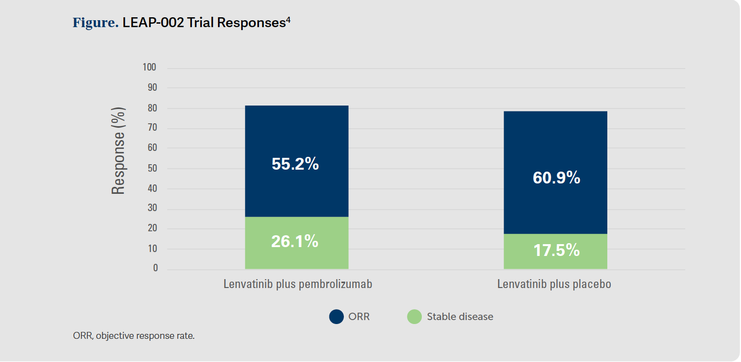
I have to say…that I find it nice to have [fewer] hand-foot-skin reactions, and I think that is less [frequent] with lenvatinib [than with] sorafenib. I do sometimes have trouble with this general fatigue, loss of appetite, and weight loss [that patients experience]. I think those sometimes can be more difficult to manage than some of the others, [and we don’t have much to treat fatigue with, which bothers lots of patients]. The hypertension and diarrhea in some cases can be tough, but in most cases they’re quite manageable if you’re monitoring [them] and tackle them early. The LEAP-002 [NCT03713593] trial data also provided some promising responses4 and AE management, as well [Figure4].


REFERENCES
1. NCCN. Clinical Practice Guidelines in Oncology. Hepatocellular carcinoma, version 1.2023. Accessed December 5, 2022. https://bit.ly/3nJuYS6
2. Management of hepatocellular carcinoma. AASLD. Accessed March 3, 2023. https://bit.ly/3V9SWlH
3. Kudo M, Finn RS, Qin S, et al. Lenvatinib versus sorafenib in first-line treatment of patients with unresectable hepatocellular carcinoma: a randomised phase 3 non-inferiority trial. Lancet. 2018;391(10126):1163- 1173. doi:10.1016/S0140-6736(18)30207-1
4. Finn RS, Kudo M, Merle P, et al. Primary results from the phase III LEAP- 002 study: lenvatinib plus pembrolizumab versus lenvatinib as first-line (1L) therapy for advanced hepatocellular carcinoma (aHCC). Ann Oncol. 2022;33(suppl 7):S1401. doi:10.1016/j.annonc.2022.08.031


Advances in Subsequent Therapies Shake Up Sequencing of ccRCC Treatment
April 25th 2024With the approval of belzutifan and other newer data for treating patients with recurrent renal cell carcinoma, the state of subsequent therapies is advancing beyond the reuse of frontline options with impacts on duration of response and quality of life.
Read More


Novel Approaches Focus on Limiting Toxicity in Older Patients With ALL
April 22nd 2024The major challenges for clinicians treating older patients with acute lymphoblastic leukemia surround the emergence of resistance to existing therapies and the toxicities associated with current chemotherapies.
Read More


Ornstein Advises on Starting Dose and Management of Lenvatinib in RCC
April 21st 2024During a Case-Based Roundtable® event, Moshe Ornstein, MD, MA, provided guidance on dosing and toxicity concerns in a patient treated with lenvatinib plus pembrolizumab for advanced renal cell carcinoma.
Read More